Upper Thigh Cross Sectional Anatomy / These horizontal planes pass through the body at right angles to the midsagittal and the frontal planes.. Atlas of body sections, ct and mri images, fourth edition. This webpage presents the anatomical structures found on orbit ct. Format_list_bulletedabout this section add remove. Computed tomography and magnetic resonance imaging. An atlas of cross sectional human anatomy.
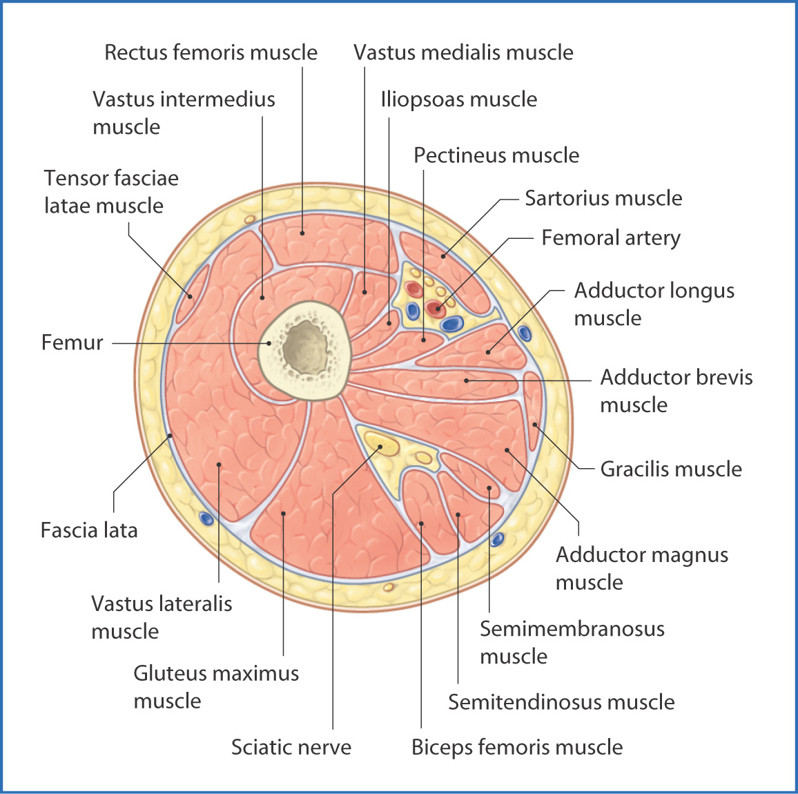
Each of these compartments has its own blood and nerve supply and contains a different group of … Chapter 15 • neuro anatomy chapter 16 • thoracic anatomy chapter 17 • abdominopelvic anatomy chapter 18 • musculoskeletal anatomy. They divide the body into upper and lower portions, and like the sagittal and frontal planes, you need to have a reference point to know exactly where a. Section 7 верхняя конечность upper limb. This webpage presents the anatomical structures found on thigh mri.
Presentation1 Pptx Radiological Anatomy Of The Thigh And Leg from image.slidesharecdn.com Mri of the upper limb. Back thigh muscles of the gluteal and posterior femoral regions from gray's anatomy of the human body from 1918. This is mainly due to the fact that the three muscle compartments (figure 6) in the thigh can compensate much higher volumes than the four compartments below the knee 1. Pelvis, perineum, hip, and upper thigh male (plates 6.1 to 6.18) female (plates 6.19 to 6.34). Format_list_bulletedabout this section add remove. Data and dicom images (archived on a pacs (picture archiving and communicating system) were processed and exported as jpeg images. There are 7 main areas covered in the upper limb; This webpage presents the anatomical structures found on orbit ct.
The arm is a region of the upper extremity located between the shoulder and elbow.
This webpage presents the anatomical structures found on thigh mri. Not very descriptive with anatomy and hard to follow. Surface anatomy is best studied using a regional. There are 7 main areas covered in the upper limb; Mri of the upper limb. The muscles, bones, joints, nerves, blood and lymphatic supply, anatomical areas, and the structures in the hand. The outer zone contains many myelinated axons that run up and down the spinal cord. Head and neck thorax abdomen upper limbs lower limbs. Skip to the end of the images gallery. Pelvis, perineum, hip, and upper thigh male (plates 6.1 to 6.18) female (plates 6.19 to 6.34). This webpage presents the anatomical structures found on orbit ct. Format_list_bulletedabout this section add remove. They divide the body into upper and lower portions, and like the sagittal and frontal planes, you need to have a reference point to know exactly where a.
Approach the regions are as follows: Exposure variables) in a population at a given point in time. Atlas of body sections, ct and mri images, fourth edition. This is mainly due to the fact that the three muscle compartments (figure 6) in the thigh can compensate much higher volumes than the four compartments below the knee 1. Skip to the end of the images gallery.
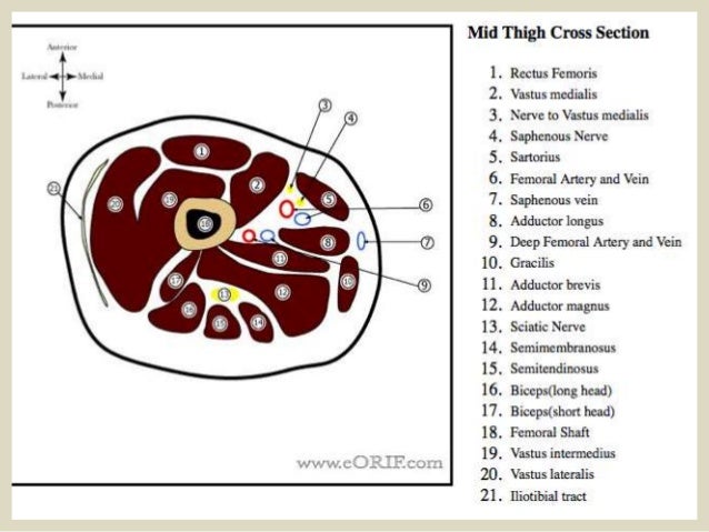
Anatomy Atlases Atlas Of Human Anatomy In Cross Section 6 Pelvis Perineum Hip And Upper Thigh from www.anatomyatlases.org Human sectional anatomy atlas of body sections, ct and mri images, fourth edition 4th edition 2015 unitedvrg.pdf. Format_list_bulletedabout this section add remove. They divide the body into upper and lower portions, and like the sagittal and frontal planes, you need to have a reference point to know exactly where a. Head and neck thorax abdomen upper limbs lower limbs. This mri brain cross sectional anatomy tool is absolutely free to use. Anatomy of the thigh and leg the thigh is best described in terms of compartmental anatomy, and is composed of anterior, posterior, and medial (adductor) compartments. Needed strictly computed tomography anatomy not mri. There are 7 main areas covered in the upper limb;
Format_list_bulletedabout this section add remove.
Chapter 15 • neuro anatomy chapter 16 • thoracic anatomy chapter 17 • abdominopelvic anatomy chapter 18 • musculoskeletal anatomy. Each of these compartments has its own blood and nerve supply and contains a different group of … Mri of the upper limb. There are 7 main areas covered in the upper limb; An atlas of cross sectional human anatomy. Format_list_bulletedabout this section add remove. Computed tomography and magnetic resonance imaging. Instant anatomy is a specialised web site for you to learn all about human anatomy of the body with diagrams, podcasts and revision questions. This is mainly due to the fact that the three muscle compartments (figure 6) in the thigh can compensate much higher volumes than the four compartments below the knee 1. Support radiopaedia and see fewer ads. Skip to the end of the images gallery. This webpage presents the anatomical structures found on thigh mri. Head and neck thorax abdomen upper limbs lower limbs.
Human sectional anatomy atlas of body sections, ct and mri images, fourth edition 4th edition 2015 unitedvrg.pdf. Mri of the upper limb. They divide the body into upper and lower portions, and like the sagittal and frontal planes, you need to have a reference point to know exactly where a. Instant anatomy is a specialised web site for you to learn all about human anatomy of the body with diagrams, podcasts and revision questions. There are 7 main areas covered in the upper limb;
Anteromedial Thigh Basicmedical Key from basicmedicalkey.com Skip to the end of the images gallery. Back thigh muscles of the gluteal and posterior femoral regions from gray's anatomy of the human body from 1918. Instant anatomy is a specialised web site for you to learn all about human anatomy of the body with diagrams, podcasts and revision questions. Lecture presentation by steven bassett southeast community college. Rhomboid muscles found in the upper back of the torso lie underneath the trapezius, providing. This webpage presents the anatomical structures found on orbit ct. The arm is a region of the upper extremity located between the shoulder and elbow. An atlas of cross sectional human anatomy.
They divide the body into upper and lower portions, and like the sagittal and frontal planes, you need to have a reference point to know exactly where a.
They divide the body into upper and lower portions, and like the sagittal and frontal planes, you need to have a reference point to know exactly where a. There are 7 main areas covered in the upper limb; These horizontal planes pass through the body at right angles to the midsagittal and the frontal planes. Femur pelvic girdle connective tissues that envelop the thigh: This is mainly due to the fact that the three muscle compartments (figure 6) in the thigh can compensate much higher volumes than the four compartments below the knee 1. Computed tomography and magnetic resonance imaging. Format_list_bulletedabout this section add remove. This webpage presents the anatomical structures found on orbit ct. Back thigh muscles of the gluteal and posterior femoral regions from gray's anatomy of the human body from 1918. Support radiopaedia and see fewer ads. Pelvis, perineum, hip, and upper thigh male (plates 6.1 to 6.18) female (plates 6.19 to 6.34). Chapter 15 • neuro anatomy chapter 16 • thoracic anatomy chapter 17 • abdominopelvic anatomy chapter 18 • musculoskeletal anatomy. Use the mouse scroll wheel to move the images up and down alternatively use the tiny arrows (>>) on both side of the image to move the images.
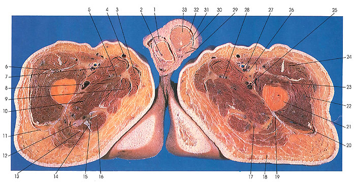
Computed tomography and magnetic resonance imaging upper thigh anatomy. Exposure variables) in a population at a given point in time.
0 Comments: